Ephydatia muelleri
Ephydatia muelleri, frequently referred to as Mueller’s Freshwater Sponge, is the most common freshwater sponge in North America. Living at the bottom of bodies of water, these poriferous creatures are immobile. These invertebrates lack organs and instead utilize complex cellular functions in order to filter water for food. In fact, their presence in a water body is an indication of high water quality, as they are quite sensitive to environmental pollution. E. muelleri is often mistaken for an alga because of its green color, however, this appearance is due to the symbiotic relationship between algae and the sponge. The sponge provides a place for the algae to live, and the algae help provide food and oxygen to the sponge.
Although the organism was considered to be a plant for hundreds of years due to its lack of mobility, further scientific inquiry revealed that sponges are quite different from the plants with which they share a habitat. Unlike a plant that has an epidermis, vascular tissue, and organs (stems, roots and leaves), the cells of E. muelleri are not organized into tissues or organs. Its specialized cells serve unique functions to keep the sponge alive including the choanocytes or “collar cells” that compose the sponge’s digestive system. The microvilli surrounding the membranes of these cells filter the surrounding water for food. Similar to plants, sponges are able to reproduce both sexually and asexually. During asexual budding, E. muelleri create gemmules, which are small structures that can withstand extreme conditions in which adult sponges cannot survive. When the environment becomes more suitable for growth the gemmule continues growing into a new individual.
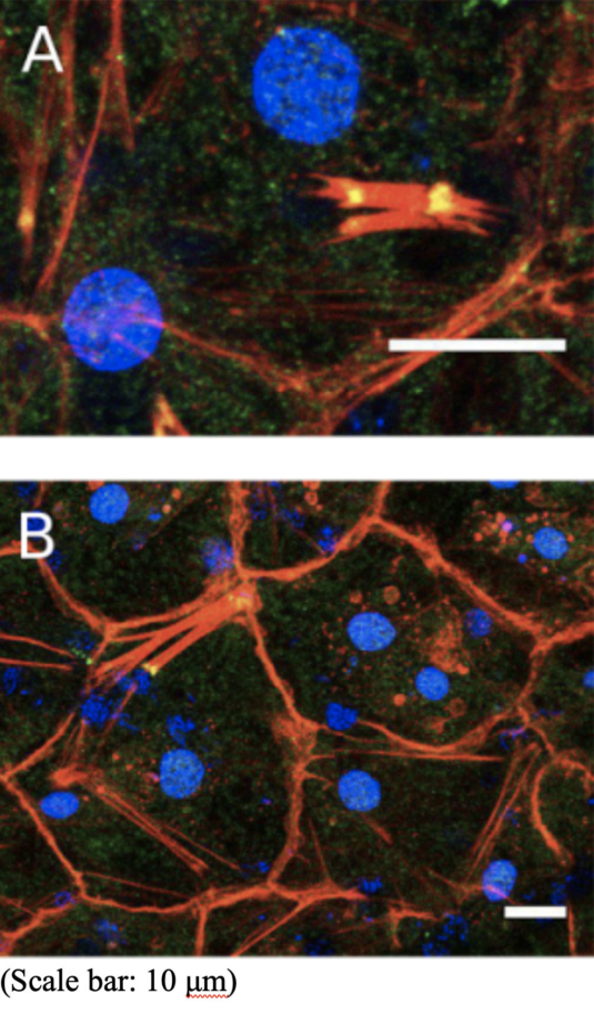
The specimen in the image above is a gemmule that was isolated from upper Red Rock Lake in the Brainard Lake Recreation Area of Colorado. The colors shown are obtained by a technique called immunofluorescence, whereby specific antibodies isolated from animals are chemically conjugated to fluorescent dyes. This antibody then attaches to another antibody that is made to affix to a molecule of interest. Next, light of a specific wavelength is shone upon the sample, and colored fluorescence of the target molecule can be observed.
The molecules of interest in this particular image are the proteins β-catenin, seen in green, actin, in red, and DNA can be seen in blue. β-catenin is a protein that is involved in transcriptional regulation and cell adhesion. Actin composes the structural cytoskeleton of the cell, and of course, DNA contains the genetic information. Together, β-catenin and actin form a complex called the adherens junction, the site where the plasma membranes of two separate cells are joined. These cell-to-cell contacts are crucial for cell communication and reducing the effects of external stress placed upon the plasma membrane.
Image source:
Schippers, K. J.; Nichols, S. (26 February 2018). “Evidence of signaling and adhesion roles for β-catenin in the sponge Ephydatia muelleri.” bioRxiv. https://doi.org/10.1101/164012

