Toxoplasma gondii
Microbestiary Photo Contest Honorable Mention
About this microbe:
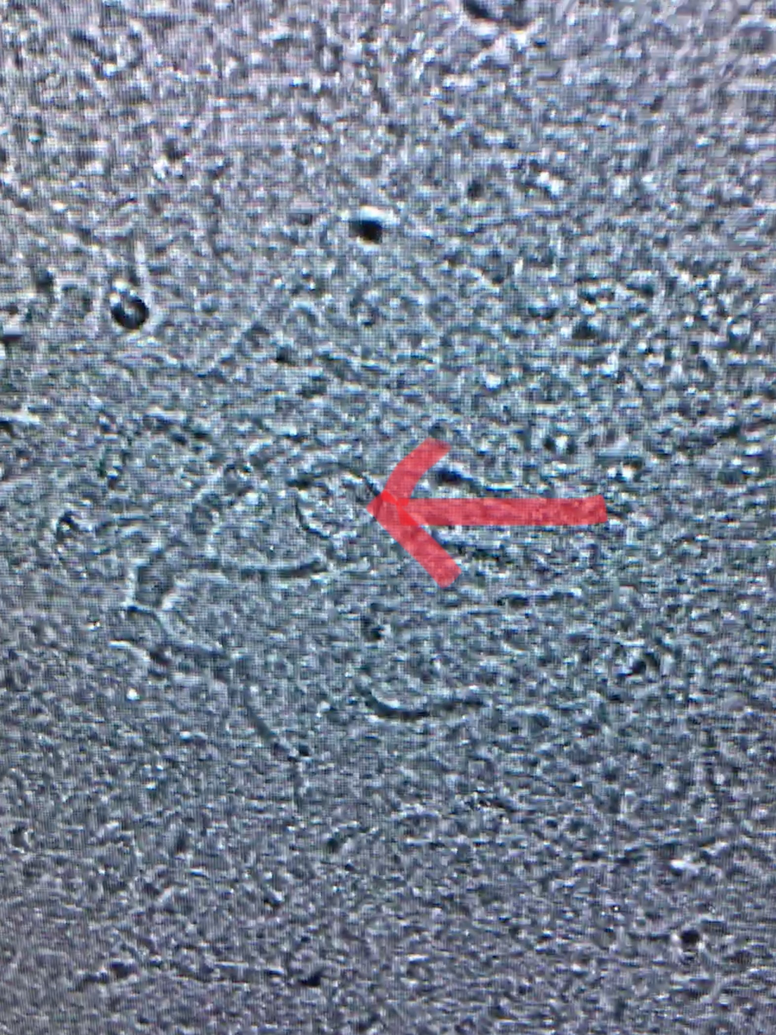
Photo 1: Toxoplasma gondii cyst seen by confocal microscope within the brain of a mouse infected for 5 weeks.
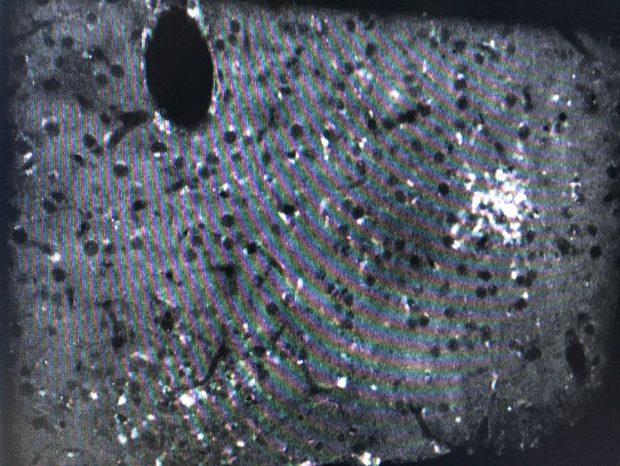
Photo 2: Cells of a mouse brain infected with Toxoplasma gondii seen under a confocal microscope. The fluorescent puncta seen are cells that are fluorescing yellow florescent protein (YFP). These cells fluoresce because they contain the NKp46 receptor and are tagged with YFP, so when they are “turned on” with Tamoxifen, their tags fluoresce yellow.
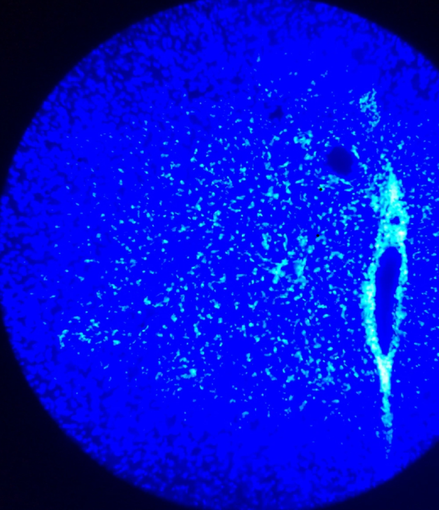
Photo 3: Cells of a mouse brain infected with Toxoplasma gondii seen under a confocal microscope. The blue seen here is because the cells are stained with DAPI staining.
Image: Sara Griffith